Determining Patterns of Failure: Advanced Imaging
How to cite: Stock, Richard G. “Determining Patterns of Failure: Advanced Imaging” April 15, 2018. Accessed [date today]. https://dev.grandroundsinurology.com/determining-patterns-of-failure-advanced-imaging/
Determining Patterns of Failure: Advanced Imaging – Summary:
Richard G. Stock, MD, discusses advanced imaging for local, nodal, and distant recurrent prostate cancer, emphasizing positron emission tomography (PET) imaging. He specifically describes the mechanisms of action and advantages of the 18F-FACBC (fluciclovine) radiotracer for PET imaging.
Patterns of Failure
Dr. Stock defines patterns of failure as recurrent disease after treatment, categorized into local, nodal, and distant. “Local” refers to recurrence in the prostate bed following prostatectomy or recurrent prostate disease after radiation therapy, brachytherapy, cryotherapy, or targeted focal therapy. “Nodal” refers to non-metastatic pelvic node recurrences, as well as para-caval/aortic recurrences. Lastly, “distant” refers to bone and visceral organ metastases.
Traditional Cancer Imaging
Traditional imaging techniques, including bone scan, computed tomography (CT) scan, and multiparametric MRI (mpMRI), are advantageous for initial staging and evaluating a patient. Regarding bone scan, sodium fluoride PET/CT scans are especially sensitive for detecting metastases in the bone. CT scan is effective for detecting nodal metastases and changes in the prostate or prostate bed. Finally, mpMRI is primarily useful for de novo disease staging.
Advanced Imaging
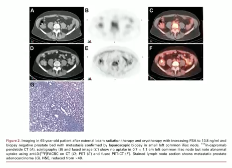
While traditional imaging is effective in early prostate cancer diagnosis, newer imaging can more accurately detect patterns of failure and recurrent disease. For example, In-111 capromab pendetide (Prostascint) single photon emission computed tomography (SPECT) shows promise in identifying recurrences in the prostate bed after prostatectomy. However, for the majority of this lecture, Dr. Stone focuses on PET imaging.
PET Imaging
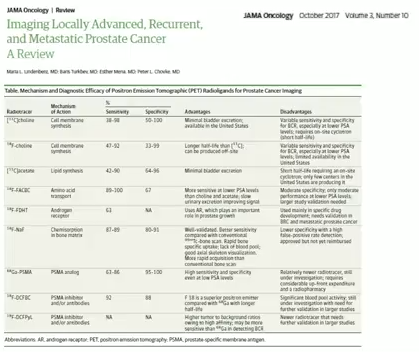
Dr. Stone briefly summarizes the mechanisms of action, advantages, and disadvantages of [11C]choline, [18F]choline, [11C]acetate, and 18F-FACBC (fluciclovine) for prostate cancer imaging. The chart below, from an article in JAMA Oncology, compares these and five additional PET radioligands.