Leonard S. Marks, MD, presented “MRI/US Fusion Biopsy” during the 24th Annual Southwest Prostate Cancer Symposium on April 13, 2019 in Scottsdale, Arizona.
How to cite: Marks, Leonard S. “MRI/US Fusion Biopsy” April 13, 2019. Accessed Apr 2024. https://dev.grandroundsinurology.com/mri-us-fusion-biopsy/
MRI/US Fusion Biopsy – Summary:
Leonard S. Marks, MD, reviews the utility of MR/US fusion prostate biopsy devices in detecting clinically significant prostate cancer and guiding targeted focal therapy. He also discusses how to develop a MR/US fusion biopsy procedure and integrate its use into a clinical practice.
Abstract:
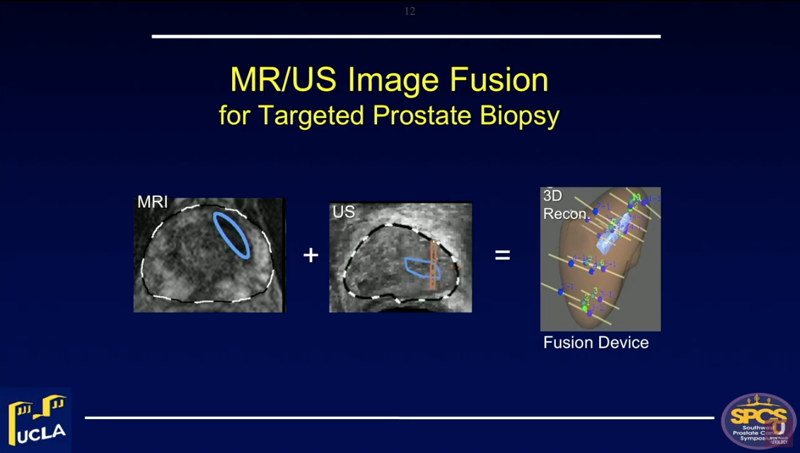
Magnetic resonance/ultrasound (MR/US) fusion prostate biopsy allows for the targeting of suspicious regions, template-mapping for systematic sampling, and tracking of cancer foci over time. All of these functions were not previously possible with US-guided biopsy.
Both the American Urological Association (AUA) and Society of Abdominal Radiology (SAR) have endorsed this procedure for men with prior negative biopsy, but continued cancer suspicion.
Targeted biopsy using MR/US fusion also improves detection of clinically significant prostate cancer (csPCa). Highly suspicious regions of interest on MRI are likely to contain cancer on targeted biopsy. The Prostate Imaging Reporting and Data System (PI-RADS) grading system of MRI is the most important determinant of biopsy outcome. Approximately 25% of PI-RADS Grade 3 lesions, or regions of interest (ROIs), will contain csPCa on biopsy. This detection rate rises to 40% with Grade 4 ROIs and to 80% with Grade 5 ROIs.
Combining targeted and systematic biopsies leads to the maximum likelihood of cancer detection. Physicians should perform both of these when performing MRI/US fusion biopsy for the first time in a man suspected of having prostate cancer. Additionally, the false negative rate of MRI is 15-20% in expert hands. Therefore, physicians should exercise caution when using a negative MRI to obviate prostate biopsy. When a biopsy is indicated clinically, a negative MRI should not preclude biopsy.
Optimally, physicians should perform biopsies using a fusion device capable of tracking biopsy site locations. This function is particularly valuable in men undergoing active surveillance for low-risk lesions. However, when both MRI is negative and PSA density is low (<0.15), the chances of finding csPCa are small (8%).
Fusion devices also provide targeting functions. Biopsy guidance, via MRI localization of cancer within the prostate, provides the foundation for focal therapies of prostate cancer, such as HIFU, cryotherapy, and laser ablation.
About the Southwest Prostate Cancer Symposium
The Southwest Prostate Cancer Symposium (SPCS) is a multi-day conference that seeks to educate urologists, radiation oncologists, medical oncologists, and other healthcare professionals involved in the treatment of prostate cancer. The topics focus on current technical aspects of diagnosis and treatment of localized and advanced disease, particularly regarding imaging, technology, and training in the related devices. Dr. Marks presented this lecture during the 24th SPCS in 2019. In 2020, the 25th SPCS will also offer training sessions involving imaging, scanning, and prostate cancer treatment related devices on site. Please visit this page in order to register for future SPCS meetings.