John F. Ward, MD, presented “MRI-US Image Fusion Prostate Biopsy” during the 24th Annual Southwest Prostate Cancer Symposium on April 13, 2019 in Scottsdale, Arizona.
How to cite: Ward, John F. “MRI-US Image Fusion Prostate Biopsy” April 13, 2019. Accessed Apr 2024. https://dev.grandroundsinurology.com/mri-us-image-fusion-prostate-biopsy/
MRI-US Image Fusion Prostate Biopsy – Summary:
John F. Ward, MD, FACS, details the workflow of the ARTEMIS Semi-Robotic Fusion Biopsy System through videos and software demonstrations. He illustrates the movement of the system’s endfire probe, the process of gathering images, rendering a digital 3D model of the prostate, and navigating the placement of biopsy needles.
Abstract:
By using the ARTEMIS Semi-Robotic Fusion Biopsy System, a multiparametric MRI/ ultrasound (MR-US) technology with semi-robotic, electronic-mechanical tracking of the prostate, clinicians can precisely navigate needles to identify lesions and significantly improve the accuracy of biopsy. This presentation provides a step-by-step walkthrough of the biopsy workflow with this system through videos and software demonstrations.
The demonstrations will cover proper maneuvering and positioning of this system’s endfire probe and application of its mechanical arm.
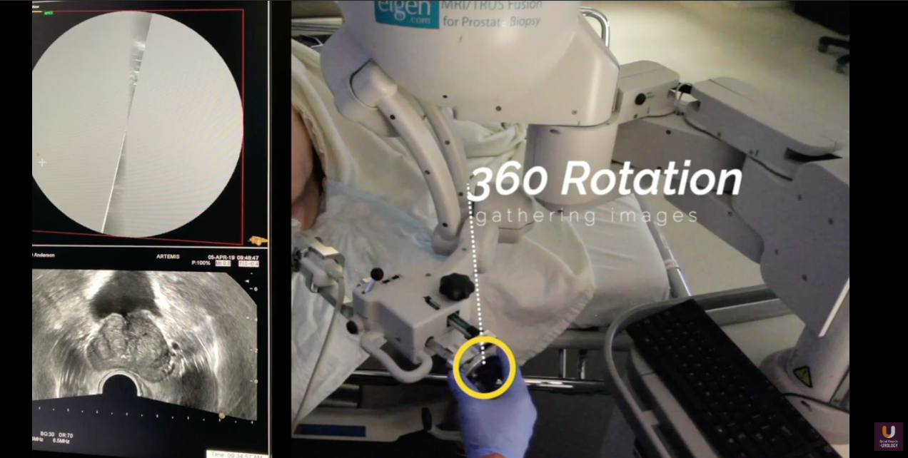
It will also illustrate the system’s utilization of 360 degrees of images gathered from the ultrasound probe and computer-generated prostate contouring, which physicians can refine manually, in order to render a 3D in-silico model of the prostate.
This system works in tandem with ProFuse Radiology Software, which similarly performs 3D modeling of the prostate through MRI, to identify, localize, and map regions of interest (ROIs). The ARTEMIS system can then align and merge this radiologic data with the ultrasound model.
This fused image and in-silico prostate model can then direct the placement of biopsy needles. The system employs motion compensation to match the previously-constructed virtual images and real-time ultrasound images while the physician navigates the needle to a target with the probe and mechanical arm. Not only can this method better target ROIs, it can also help avoid under- and oversampling in systematic biopsies. The system will record the history of exact locations of cores taken, as well. Finally, the system has the ability to import previously-recorded information from a patient, which is beneficial for future biopsies.
About the Southwest Prostate Cancer Symposium
The Southwest Prostate Cancer Symposium (SPCS) is a multi-day conference that seeks to educate urologists, radiation oncologists, medical oncologists, and other healthcare professionals involved in the treatment of prostate cancer. The topics focus on current technical aspects of diagnosis and treatment of localized and advanced disease, particularly regarding imaging, technology, and training in the related devices. Dr. Ward presented this lecture during the 24th SPCS in 2019. In 2020, the 25th SPCS will also offer training sessions involving imaging, scanning, and prostate cancer treatment related devices on site. Please visit this page in order to register for future SPCS meetings.